
Finding a new bump while stroking your Frenchie can send you straight into a photo-search spiral. If I found a new bump on Nina, I would put that energy into a clearer next step: tell your vet what you found and arrange an examination. A photograph, a soft texture or a familiar-looking skin tag cannot reliably tell you what a new growth is.
This guide is about a distinct lump on or beneath the skin. If the main change is widespread itching rather than one growth, our French bulldog allergy guide covers that different starting point. Neither pattern is a diagnosis by itself.
The useful takeaways
- Have a new lump assessed, even if a previous one was harmless.
- Keep a location record so you and your vet discuss the same bump.
- A needle sample and a tissue biopsy answer related but different questions.
- An inconclusive sample is not an all-clear. Ask what happens next.
Describe the change without trying to name it
“A bump behind her left shoulder” is more useful than “probably a cyst.” A vet may consider inflammation, a cyst, a benign growth or a malignant tumour, among other possibilities. The 2026 AAHA oncology guidelines explain that the nature and likely behaviour of a mass cannot be established from feeling it or imaging alone. Microscopic assessment is an important part of investigating a mass.
That does not mean every bump is cancer. It means you do not have to solve that question with your fingers. Avoid repeatedly pressing, picking or squeezing the area to see what comes out.
Make a small record before the appointment
The PDSA’s veterinary guidance on skin lumps recommends contacting your vet about each new lump and documenting its size and appearance. Tell the clinic if it is growing, bleeding, leaking, bothering your dog or affecting movement; these changes can affect how soon they want to see your pet.
A simple phone note keeps the conversation organised:
- Location: describe the side and nearby landmark. Give separate bumps separate labels.
- First noticed: record the date you found it, not an assumed date it began.
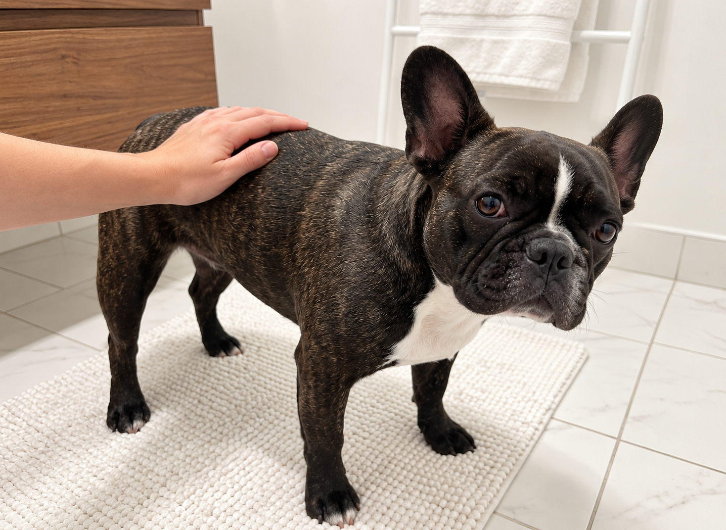
- Photo: take an overview that shows the location, plus a closer picture if your dog is comfortable.
- Changes: note what you actually saw, including licking, discharge or a change in daily activity.
- Previous result: bring any report for that particular site, rather than relying on “she has had lumps before.”
If your dog dislikes handling, stop. A useful note does not require restraining her for a perfect photograph. Photos help communication; they do not replace the examination.
What a needle sample can tell you
Fine-needle aspiration, often shortened to FNA, collects cells for examination under a microscope. Your vet may review them or send them to a laboratory. VCA’s explanation of FNA describes both its usefulness and its limits: sometimes too few cells are collected, or the cells on the slide do not represent the whole lesion.
Ask two separate questions when results arrive: “What did the sample show?” and “Was it sufficient to answer the question about this lump?” An answer such as “not enough diagnostic cells” means there is still uncertainty. Discuss whether another sample, a different sampling method or a biopsy is appropriate. Do not translate an inconclusive report into either “definitely benign” or “definitely cancer.”
Why a biopsy may be the next step
A biopsy supplies tissue rather than just individual cells. According to AAHA, examining tissue can reveal information that cell samples may not provide, including architecture, invasiveness and, for relevant tumours, grade. Your vet should explain how the proposed test would change the plan.
Useful questions include: Will you sample part of the lump or remove it? Will the tissue go for laboratory examination? What comfort, sedation or anaesthetic plan is needed for my dog? Who will contact me with the result, and when should I call if I have not heard?
Monitoring should be a specific plan
MSPCA-Angell’s clinical guidance explains why appearance alone can mislead: an irritated growth can be benign, while a quiet, slow-growing one can be malignant. Management after assessment may range from observation to treatment. A benign lump can still need attention if it causes discomfort or interferes with movement.
If monitoring is advised, ask which lump that instruction applies to, what changes should trigger a call, and when it should be checked again. I would keep the result and the follow-up instructions in the same note as the location photo. “Watch it” is much easier to follow when everyone means the same thing.
Common questions
Can I remove a skin tag myself?
No. Have the growth identified and discuss whether it needs treatment. Do not cut, tie off or use human wart-removal products on it.
Does every lump need surgery?
No. The decision depends on the diagnosis, location, behaviour and effect on your dog. The first goal is a useful assessment, not automatically an operation.
Can I assume another soft bump is the same as an old one?
No. Report the new site separately. A reassuring result belongs to the lump that was assessed, not to every future bump.
This is owner education, not a diagnosis of your dog’s skin. Your veterinary team should direct testing, treatment and follow-up.